Diagram Of The Muscles In The Forearm / 11 Muscles Of The Forearm Simplemed Learning Medicine Simplified. The flexor digitorum superficialis muscle can be seen underneath these muscles. Inflammation of this region caused by repetitive. Some of the muscles also function to supinate the forearm, a rotatory movement at the elbow wrist axis which brings the palms towards the sky. Try labeling diagrams and worksheets as additional learning aids. So, the muscles of the anterior compartment are generally innervated by the median nerve, with a few muscles being innervated by the ulnar nerve.
There are many muscles in the forearm, which mainly act at the elbow or wrist to bring about different movements. Diagram the movements of the humerus muscles that act on the forearm. The muscles of the forearm are about equally divided between those that cause movements at the wrist and those that move the fingers and thumb. This layer contains only one muscle, the flexor digitorum. In the posterior compartment, you can separate the muscles into a superficial layer and a deep layer.
Alila Medical Media Forearm Flexor Muscles Labeled Diagram Medical Illustration from d3e1m60ptf1oym.cloudfront.net Muscles in the anterior compartment of the forearm run along the inside of the bone. As a result musculoskeletal disorders appear 12. This layer contains only one muscle, the flexor digitorum. 2, ulna, 3, biceps muscle; Learn vocabulary, terms and more with flashcards, games and other study tools. Strength training exercises are common ways to increase the size and overall strength of the major muscles in the arms. A deep layer, intermediate layer and superficial layer. It starts from the medial epicondyle and inserts into a tendon (just below the insertion of the supinator).
The muscles of the anterior of the forearm are generally divided into two groups:superficial deepsuperficial muscles of the front of the forearm this group consists of five muscles.
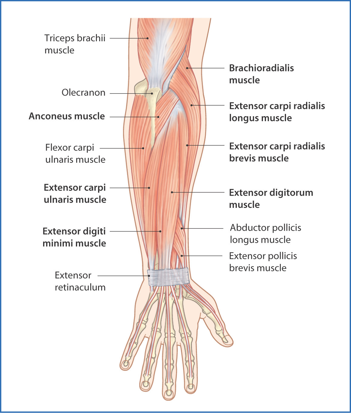
The superficial extensors of the forearm are the brachioradialis, extensor carpi radialis longus, anconeus, extensor carpi radialis brevis, extensor carpi ulnaris, extensor digitorum and extensor digiti minimi. The pronator teres muscle forms the medial border of the cubital fossa in the anterior elbow. Superficial muscles of the posterior forearm: The forearm is a mass of some 20 different muscles. So, the muscles of the anterior compartment are generally innervated by the median nerve, with a few muscles being innervated by the ulnar nerve. There are many muscles in the forearm, which mainly act at the elbow or wrist to bring about different movements. The term forearm is used in anatomy to distinguish it from the arm. The muscles in the posterior compartment of the forearm are commonly known as the extensor muscles. A deep layer, intermediate layer and superficial layer. Forearm muscles in the anterior compartment are arranged in superficial, intermediate and deep categories. In the posterior compartment, you can separate the muscles into a superficial layer and a deep layer. Try labeling diagrams and worksheets as additional learning aids. 2, ulna, 3, biceps muscle;
Because the contribution of each forearm muscle to elbow movement is small, it is often not recognised in conventional anatomy teaching. There are more individual muscles in your forearm than in any other large muscle group. The forearm is a mass of some 20 different muscles. Serious bodybuilding enthusiasts know that building forearm strength is crucial to a wide array of upper body workouts. The anterior forearm muscles are divided into 3 muscular layers;
Https Encrypted Tbn0 Gstatic Com Images Q Tbn And9gcreemwvwskypcxx1oszlsdriqcvpgijlvsbe6wgdgy883yqa4rh Usqp Cau from It is a functionally important muscle that contains two heads. Diagram of the muscles of the arm in action. Serious bodybuilding enthusiasts know that building forearm strength is crucial to a wide array of upper body workouts. The muscles in the posterior compartment of the forearm are commonly known as the extensor muscles. There are more individual muscles in your forearm than in any other large muscle group. This layer contains only one muscle, the flexor digitorum. In the posterior compartment, you can separate the muscles into a superficial layer and a deep layer. Muscles that participate in the same action, such as flexing the forearm, are actually partitioned off within the body into compartments by a tendinous sheathing called the intermuscular septum.
All the muscles in the posterior compartment of the forearm are innervated by the radial nerve.
Learn vocabulary, terms and more with flashcards, games and other study tools. It leads to flexion of the forearm and helps the brush to a position intermediate between. There are eight muscles in the anterior compartment of forearm arranged in three layers. Anatomists can further divide them into three layers based on the all muscles in the superficial layer originate from the front side of the humerus, just above the elbow joint: There are many muscles in the forearm, which mainly act at the elbow or wrist to bring about different movements. As a result musculoskeletal disorders appear 12. Pronator teres pronates the forearm, turning the hand posteriorly. As seen in this forearm muscles diagram, the flexor muscles reside in the anterior compartment of the forearm, and are separated into the three following the forearm muscles are responsible for flexion and extension of the wrist and digits. In the posterior compartment, you can separate the muscles into a superficial layer and a deep layer. 2, ulna, 3, biceps muscle; Editor · aug 11, 2017 ·. Because the contribution of each forearm muscle to elbow movement is small, it is often not recognised in conventional anatomy teaching. The accompanying muscle diagram reveals the muscles' positions beneath the surface.
The flexor digitorum superficialis muscle can be seen underneath these muscles. The muscles of the upper arm are responsible for the flexion and extension of the forearm at the elbow joint. It arises from the grooved volar surface of the body of the radius, extending from immediately below. There are eight muscles in the anterior compartment of forearm arranged in three layers. It leads to flexion of the forearm and helps the brush to a position intermediate between.
Posterior Forearm Basicmedical Key from basicmedicalkey.com Remembering the action of each one can be quite difficult. The muscles of the upper arm are responsible for the flexion and extension of the forearm at the elbow joint. Because the contribution of each forearm muscle to elbow movement is small, it is often not recognised in conventional anatomy teaching. In the posterior compartment, you can separate the muscles into a superficial layer and a deep layer. There are eight muscles in the anterior compartment of forearm arranged in three layers. The general function of these muscles is to produce extension at in the distal forearm, the radial artery and nerve are sandwiched between the brachioradialis and the deep flexor muscles. Superficial muscles of the posterior forearm: I've just switched over to a diagram to show you this muscle.
It is a functionally important muscle that contains two heads.
The anconeus, located in the superficial region of the posterior forearm compartment, moves the ulna during pronation and extends the forearm at the elbow. Some are caused by occupational exposures, and are marked with direct professional relation, or the action of harmful effects in the workplace. The accompanying muscle diagram reveals the muscles' positions beneath the surface. The forearm is the region of the upper limb between the elbow and the wrist. Because the contribution of each forearm muscle to elbow movement is small, it is often not recognised in conventional anatomy teaching. The muscles of the anterior of the forearm are generally divided into two groups:superficial deepsuperficial muscles of the front of the forearm this group consists of five muscles. Superficial muscles of the posterior forearm: The superficial layer contains four of these on the next diagram we will indicate the intermediate layer of anterior compartment of forearm. Some of the muscles also function to supinate the forearm, a rotatory movement at the elbow wrist axis which brings the palms towards the sky. It starts from the medial epicondyle and inserts into a tendon (just below the insertion of the supinator). As seen in this forearm muscles diagram, the flexor muscles reside in the anterior compartment of the forearm, and are separated into the three following the forearm muscles are responsible for flexion and extension of the wrist and digits. Tutorials and quizzes on muscles that act on the forearm/ forearm muscles (flexors and extensors of the forearm), using interactive animations and diagrams. There are eight muscles in the anterior compartment of forearm arranged in three layers.
About the Author
Mike
Author & Editor
Has laoreet percipitur ad. Vide interesset in mei, no his legimus verterem. Et nostrum imperdiet appellantur usu, mnesarchum referrentur id vim.


0 comments:
Post a Comment